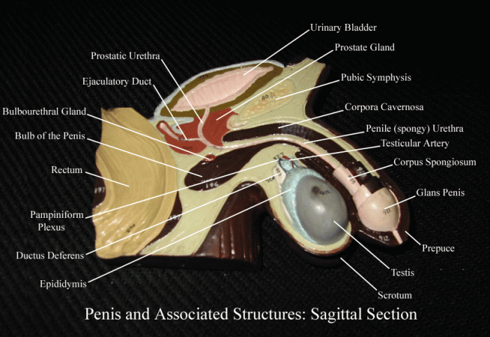
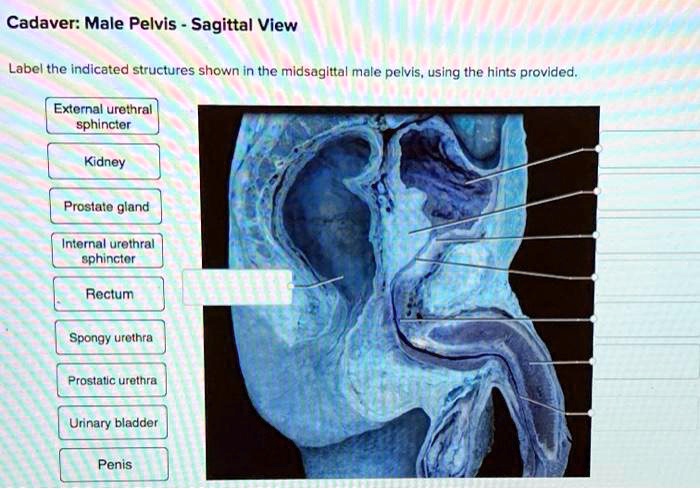
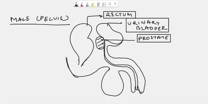
Label the midsagittal male pelvis using the hints provided. – Labeling the midsagittal male pelvis using the hints provided requires a comprehensive understanding of the pelvic structures and their anatomical relationships. This guide provides a detailed overview of the male pelvis, including the os coxae, sacrum, coccyx, pubis, ischium, ilium, pelvic inlet, pelvic outlet, and pelvic floor muscles.
By understanding the anatomy of the male pelvis, healthcare professionals can better diagnose and treat pelvic disorders.
The male pelvis is a complex and dynamic structure that plays a crucial role in various bodily functions, including reproduction, urination, and defecation. The accurate labeling of the midsagittal male pelvis is essential for medical professionals to effectively communicate and collaborate in patient care.
Label the structures of the male pelvis using hints

The male pelvis is a bony structure that forms the lower part of the trunk. It is composed of four bones: the two hip bones (os coxae), the sacrum, and the coccyx. The hip bones are large, flat bones that form the sides and front of the pelvis.
Each hip bone is composed of three parts: the ilium, the ischium, and the pubis. The sacrum is a triangular bone that forms the back of the pelvis. It is composed of five fused vertebrae. The coccyx is a small, triangular bone that forms the tip of the pelvis.
It is composed of four fused vertebrae.
Location of the os coxae and its components
The os coxae is located on either side of the pelvis. It is composed of three parts: the ilium, the ischium, and the pubis. The ilium is the largest part of the os coxae. It forms the upper part of the pelvis and flares out laterally to form the iliac crest.
The ischium is located below the ilium. It forms the lower and back part of the pelvis and has a large, curved projection called the ischial tuberosity. The pubis is located below the ischium. It forms the front of the pelvis and has a large, flat projection called the pubic bone.
Details on the sacrum and coccyx
The sacrum is a triangular bone that forms the back of the pelvis. It is composed of five fused vertebrae. The sacrum is curved forward and has a concave anterior surface and a convex posterior surface. The coccyx is a small, triangular bone that forms the tip of the pelvis.
It is composed of four fused vertebrae. The coccyx is curved backward and has a concave anterior surface and a convex posterior surface.
Structure and position of the pubis, ischium, and ilium, Label the midsagittal male pelvis using the hints provided.
The pubis is located below the ischium. It forms the front of the pelvis and has a large, flat projection called the pubic bone. The pubic bone is connected to the ischium by the pubic symphysis. The ischium is located below the ilium.
It forms the lower and back part of the pelvis and has a large, curved projection called the ischial tuberosity. The ischial tuberosity is the point of attachment for the hamstring muscles. The ilium is the largest part of the os coxae.
It forms the upper part of the pelvis and flares out laterally to form the iliac crest. The iliac crest is the point of attachment for the abdominal muscles.
Identify the pelvic inlet and outlet

Definition of the pelvic inlet and its boundaries
The pelvic inlet is the superior opening of the pelvis. It is bounded by the pubic bones anteriorly, the ilia laterally, and the sacrum posteriorly. The pelvic inlet is a heart-shaped opening that is wider at the sides than at the front or back.
Shape and dimensions of the pelvic outlet
The pelvic outlet is the inferior opening of the pelvis. It is bounded by the pubic bones anteriorly, the ischia laterally, and the sacrum and coccyx posteriorly. The pelvic outlet is a diamond-shaped opening that is wider at the back than at the front.
The pelvic outlet is smaller than the pelvic inlet.
Clinical significance of the pelvic inlet and outlet
The pelvic inlet and outlet are important clinical landmarks. The size and shape of the pelvic inlet and outlet can affect the course of labor and delivery. A narrow pelvic inlet or outlet can make it difficult for the baby to pass through the birth canal.
This can lead to complications such as prolonged labor, fetal distress, and cesarean delivery.
Describe the pelvic floor muscles

List of muscles that form the pelvic floor
The pelvic floor muscles are a group of muscles that form the floor of the pelvis. They include the levator ani muscle, the coccygeus muscle, and the pubococcygeus muscle.
Function of each muscle
- The levator ani muscle supports the pelvic organs and helps to control urination and defecation.
- The coccygeus muscle supports the pelvic organs and helps to control defecation.
- The pubococcygeus muscle supports the pelvic organs and helps to control urination.
Clinical implications of pelvic floor dysfunction
Pelvic floor dysfunction can occur when the pelvic floor muscles are weakened or damaged. This can lead to a variety of symptoms, including urinary incontinence, fecal incontinence, and pelvic organ prolapse. Pelvic floor dysfunction can be treated with a variety of methods, including pelvic floor exercises, electrical stimulation, and surgery.
Compare the male and female pelvis
Key anatomical differences between the male and female pelvis
- The male pelvis is narrower and deeper than the female pelvis.
- The male pelvic inlet is heart-shaped, while the female pelvic inlet is oval-shaped.
- The male pelvic outlet is smaller than the female pelvic outlet.
- The male sacrum is narrower and more curved than the female sacrum.
- The male coccyx is shorter and less curved than the female coccyx.
Functional significance of these differences
The anatomical differences between the male and female pelvis are related to the different functions of the pelvis in each sex. The male pelvis is designed to support the weight of the upper body and to protect the pelvic organs.
The female pelvis is designed to allow for childbirth. The wider and shallower female pelvis allows the baby to pass through the birth canal more easily.
Implications for childbirth and other pelvic functions
The differences between the male and female pelvis have implications for childbirth and other pelvic functions. The narrower male pelvis can make it more difficult for the baby to pass through the birth canal. This can lead to complications such as prolonged labor, fetal distress, and cesarean delivery.
The wider female pelvis allows the baby to pass through the birth canal more easily. This makes childbirth less likely to be complicated.
Illustrate the pelvic structures using medical imaging

Examples of medical imaging techniques used to visualize the pelvis
There are a variety of medical imaging techniques that can be used to visualize the pelvis. These include X-rays, computed tomography (CT) scans, and magnetic resonance imaging (MRI) scans.
How these techniques can be used to diagnose and treat pelvic disorders
Medical imaging techniques can be used to diagnose and treat a variety of pelvic disorders. For example, X-rays can be used to diagnose fractures of the pelvic bones. CT scans can be used to diagnose tumors and other abnormalities of the pelvic organs.
MRI scans can be used to diagnose soft tissue injuries of the pelvis.
Advantages and disadvantages of different imaging modalities
Each medical imaging technique has its own advantages and disadvantages. X-rays are relatively inexpensive and widely available. However, they can only provide two-dimensional images of the pelvis. CT scans provide more detailed images than X-rays, but they are more expensive and involve exposure to radiation.
MRI scans provide the most detailed images of the pelvis, but they are the most expensive and time-consuming.
Top FAQs: Label The Midsagittal Male Pelvis Using The Hints Provided.
What are the components of the os coxae?
The os coxae consists of the ilium, ischium, and pubis.
What is the shape of the pelvic inlet?
The pelvic inlet is heart-shaped.
What muscles form the pelvic floor?
The pelvic floor muscles include the levator ani, coccygeus, and pubococcygeus muscles.